

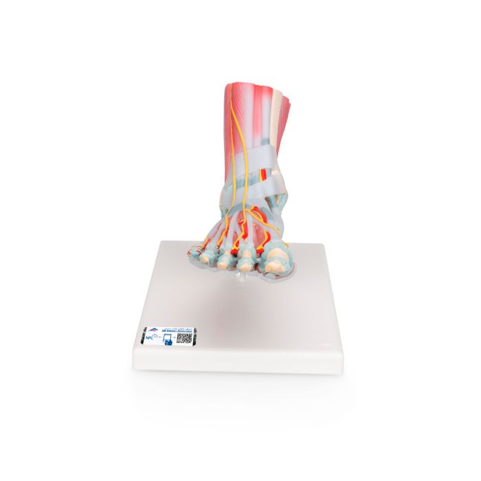
3B Scientific Foot Skeleton Model with Ligaments and Muscles

Grades 6–12 Examine the human foot in great detail with this anatomically correct model. The bones, muscles, tendons, ligaments, nerves, arteries, and veins are on full display in this six-part model of the foot and ankle.
The frontal view of the foot model features the extensor muscles of the lower leg. The tendons can be followed on their passage under the transverse and crucial crural ligaments all the way to their insertion points. On the dorsal portion of the foot, the gastrocnemius muscle is removable so students can study the deeper anatomical elements. The sole of the foot is represented in three layers. The first layer displays the flexor digitorum brevis muscle, which can be removed from the model to reveal the quadratus plantae, the tendon of the flexor digitorum longus, and the flexor hallucis muscle.
This high-quality model includes free access to the anatomy course 3B Smart Anatomy, which includes 23 digital anatomy lectures, 117 different virtual anatomy models, and 39 anatomy quizzes to test student knowledge using the Complete Anatomy app by 3D4Medical. Allow extra delivery time.


| Brand | American 3B Scientific |
|---|---|
| Item Weight | 4 lbs |
| Length | 14.00 |
| Width | 7.00 |
| Height | 9.00 |
| Age | 11 yrs-18 yrs |
| Grade | 6-12 |
| Prop 65 |
|
| School Type | High School, Middle School |

-
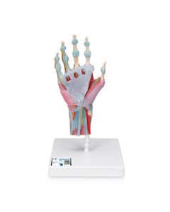 3B Scientific Hand Skeleton Model with Ligaments and Muscles $424.32Product Number: NE40265In Stock